
Common conditions

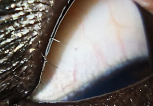
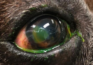
Nictitans Gland Prolapse – ‘Cherry Eye’

Cherry eye refers to prolapse of the nictitans gland and is seen more commonly in dogs compared to cats. It can occur in any breed, however brachycephalic (flat-faced) breeds, mastiffs and Great Danes are over-represented. Either one or both eyes can be affected and the condition is most commonly seen in young dogs.

Corneal sequestrum

Corneal sequestra are unique to cats and are pigmented lesions that develop on the surface of the eye. They can range from light brown ‘tea-stain’ like lesions to dark, scab like lesions and in some cases can cause significant discomfort.

Deep corneal ulceration

Deep ulcers are those which involve the deeper layers of the cornea beneath the epithelium, known as the corneal stroma. A previously ‘simple’ ulcer caused by dry eye, eyelid abnormalities or trauma for example can become deep if the underlying cause remains untreated or if it becomes infected.

Distichiasis

Distichia are hairs which grow abnormally from glands on the eyelid margin rather than from hair follicles on the skin. This means that they contact the surface of the eye and can be very irritating.

Entropion

Entropion is a condition where one or both eyelids roll inwards. This not only irritates the surface of the eye but can also result in ‘trichiasis‘. This is when hairs from the eyelid skin contact the surface of the eye causing further irritation and damage.

Primary Closed-Angle Glaucoma – ‘Goniodysgenesis’

Glaucoma refers to increased pressure within the eye. In humans glaucoma is described as a migraine-like pain. We assume it is also painful in our animals given the improvement in their demeanour following treatment.

Lens luxation

The lens is situated behind the iris and is suspended in place by numerous small fibres called lens zonules. Lens luxation refers to dislocation of the lens within the eye and can either be anterior (shifts forward) or posterior (shifts backwards).

Melting ulcers (Keratomalacia)

A melting ulcer, or keratomalacia, occurs when the cornea starts to lose its solid structure and becomes ‘jelly’ like. They can rapidly progress to rupture of the eye within 24 hours and are therefore an ocular emergency.

Sudden acquired retinal degeneration syndrome (SARDS)

SARDS occurs when the number of light-receiving cells (photoreceptors) within the retina rapidly reduce in number. Numerous studies have been performed to determine the cause of this loss, however as yet the cause remains unknown and research is ongoing.

SCCED (Spontaneous chronic corneal epithelial defect)

SCCEDs are a type of corneal ulcer that are unique to dogs and can be very slow to heal, making treatment very frustrating for vets and owners. They occur spontaneously in any breed and are more common in older dogs. They form when the top layer of corneal cells (epithelium) does not adhere to the underlying collagen, giving them a characteristic ‘lip’ of loose corneal tissue around the edge of the ulcer.

Quantitative dry eye (Keratoconjunctivitis sicca)

Quantitative dry eye occurs due to a reduced production of tears. The most common cause in dogs is autoimmune disease, where the immune system inappropriately attacks the lacrimal gland which is responsible for the majority of tear production.

Dogs are usually middle-aged when dry eye begins and although any dog can be affected, certain breeds such as the English bulldog, West Highland white terrier and spaniel breeds are more commonly represented.

Uveitis

Uveitis refers to inflammation of structures within the eye and can be further classified as: 1) Anterior uveitis: affecting structures in the front of the eye such as the iris. 2) Posterior uveitis: affecting structures in the back of the eye such as the retina. 3) Panuveitis: concurrent anterior and posterior uveitis.